Pet Digital Dental X-Rays in Tucson, AZ
Oral health plays a central role in your pet’s overall well-being. Many dental problems begin below the gum line, where they can’t be seen during a standard exam. Cimarron Animal Hospital offers pet digital dental X-rays in Tucson, AZ, to provide accurate, detailed evaluations of your pet’s teeth and surrounding structures. This diagnostic tool allows our team to detect hidden conditions early and plan appropriate treatment before discomfort worsens.
Why Digital Dental X-Rays Are Essential in Veterinary Dentistry
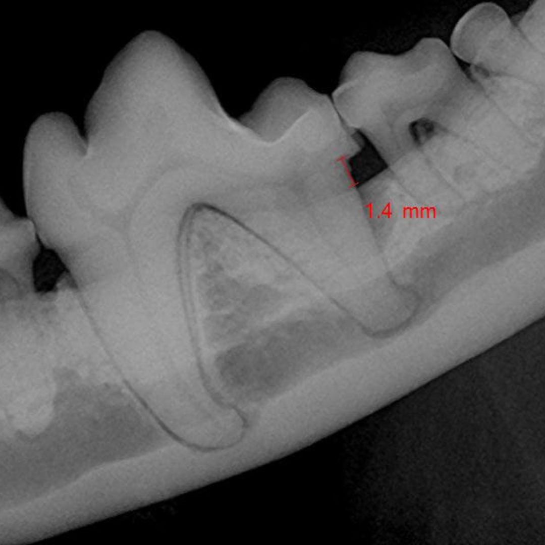
While physical exams help identify tartar buildup and damaged teeth, they can’t show what’s happening beneath the surface. Digital dental X-rays allow veterinarians to evaluate the roots, jawbone, and tissues below the gum line. These high-resolution images are a vital part of diagnosing and treating oral health issues in both dogs and cats.
Conditions often detected using pet digital dental x-rays include:
- Root infections that may not cause visible signs but can lead to pain or tooth loss
- Jawbone loss related to advanced periodontal disease
- Oral tumors or masses affecting tooth roots and nearby tissues
- Tooth fractures that require extraction or ongoing monitoring
- Resorptive lesions in cats, a painful condition best diagnosed through imaging
By identifying these issues early, your pet can receive targeted treatment that prevents complications and supports long-term health.
How Cimarron Animal Hospital Uses Pet Digital Dental X-Rays
Cimarron Animal Hospital includes digital X-rays as part of every comprehensive dental procedure. The process is safe, quick, and non-invasive. Pets are placed under general anesthesia for thorough cleaning and imaging, minimizing stress and movement during the procedure.
Once the X-rays are taken, our team reviews them to assess tooth and bone health. Based on the results, we can determine the best course of action—whether that involves cleaning, extraction, or follow-up monitoring.
Key Advantages of Pet Digital Dental X-Rays
Digital radiography improves both diagnosis and treatment planning. The benefits include:
- Improved accuracy through clear, detailed images
- Better treatment decisions by confirming whether a tooth is salvageable or needs removal
- Early detection of hidden infections and oral diseases
- Ongoing monitoring of dental health over time
Each X-ray provides the information needed to support your pet’s comfort, function, and quality of life.
What to Expect During Your Pet’s Dental Visit
When your pet is scheduled for a dental procedure at Cimarron Animal Hospital, digital X-rays are part of the care plan. These radiographs are reviewed in real time to guide any necessary treatments during the same visit.
Our veterinary team explains findings clearly and discusses recommended next steps, ensuring you’re informed at every stage. Follow-up care may be advised depending on what is revealed in the imaging and how your pet’s mouth responds to treatment.
Book a Dental Assessment Today
Oral health issues can develop quietly, and by the time symptoms appear, the problem may already be advanced. Pet digital dental X-rays in Tucson, AZ, help detect these issues early, providing your pet with a better chance at lasting dental health.
Contact Cimarron Animal Hospital today to book your pet’s dental appointment. Our team is here to support comprehensive care with the diagnostic tools needed to keep your pet comfortable and thriving.